Products
MRMath provides industry-leading products designed to impact medical imaging.
View our advanced innovative tools and services for healthcare professionals
and patients.
-Smart Manual Segmentation
The MRIMath manual contouring platform uses advanced mathematical tools to ensure high efficiency and consistency, with inter-user variability below 5%.
Alongside its intelligent features, the interface includes essential tools for drawing, erasing, painting, editing, adjusting borders, and interpolation — all designed for ease of use.

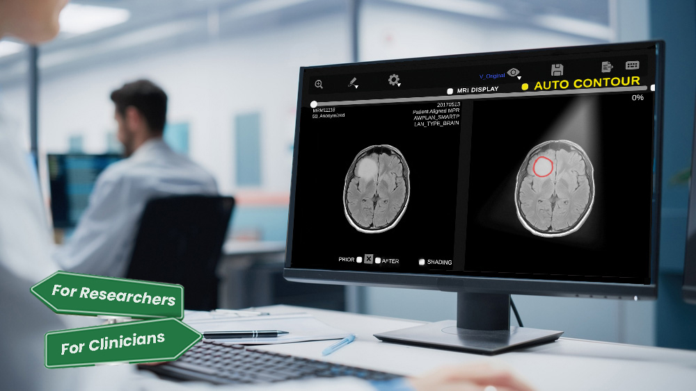
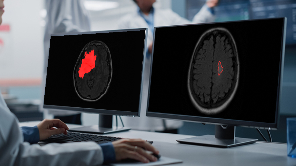
-AI-Powered Segmentations
High-performance AI segmentation — designed for speed, accuracy, and clinical impact.
Our AI-powered platform accelerates the process, with typical segmentation and review completed in under 2–3 minutes — up to 30× faster than manual methods.
This efficiency helps reduce delays across the entire workflow, without sacrificing precision.
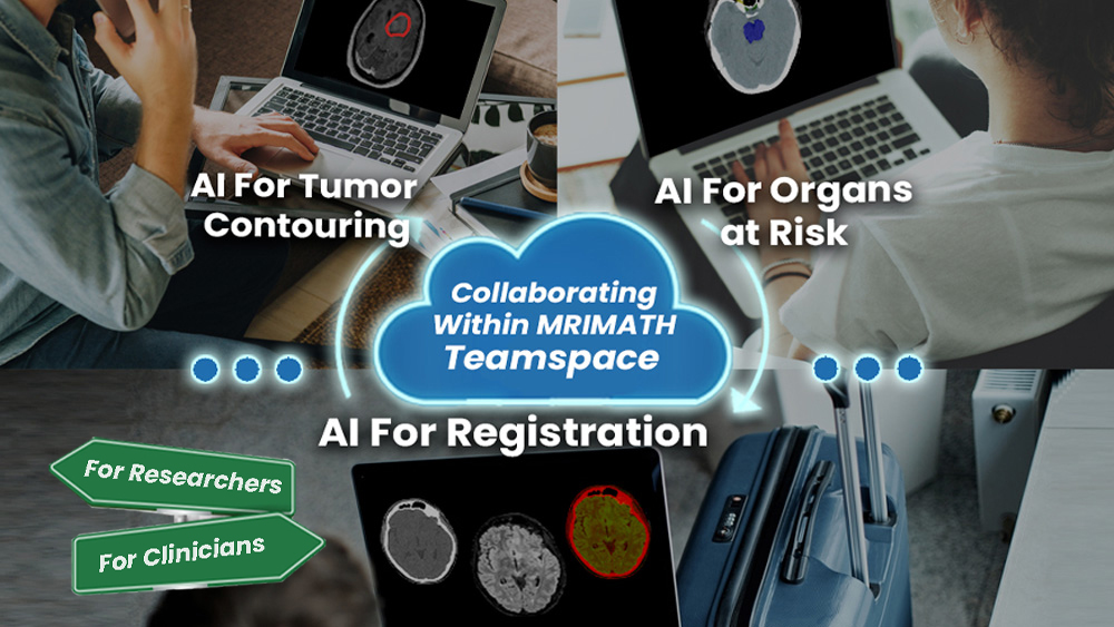
MRIMath solutions operate seamlessly within a unified teamspace to address critical clinical bottlenecks, including both tumor and OAR segmentation.
We currently offer:
- AI-driven GBM segmentation (FDA approved)
- AI-driven GBM segmentation with uncertainty quantification (FDA in preparation)
- AI-driven metastatic brain tumor segmentation (FDA in preparation)
- AI-driven organs-at-risk segmentation for the brain (FDA in preparation)
-Collaborative TeamSpace
Real-time collaboration. Seamless productivity.
Whether you’re working across the hall or across the globe, our TeamSpace keeps everyone connected — securely and in real time.
Key features include:
- Accelerated workflows and streamlined task management
- Full control over your data and permissions
- Easy team onboarding and role assignments
- Seamless collaboration on patient care or research
- Scalable access for multi-institutional teams
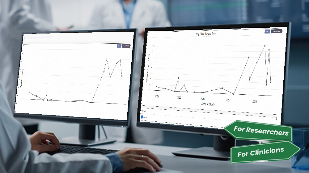
-GBM Volumetric Measurements/Plots
Track Tumor Progression with Precision.
Quantify change. Visualize impact. Guide treatment.MRIMath enables clinicians and researchers to monitor glioblastoma (GBM) tumor volumes over time with clarity and consistency.
Our platform transforms longitudinal data into intuitive, high-resolution plots—helping teams detect meaningful shifts in tumor growth and confidently assess treatment response.
Use volumetric trends to:
- Compare pre- and post-treatment tumor volume (FDA approved)
- Measure percentage change over time (FDA approved)
- Detect statistically significant changes using advanced analytics (FDA in preparation)
-Cyber Device With Secure Communications
Work securely — from anywhere.
MRIMath’s platform is built with government-grade cybersecurity, meeting the standards required for FDA clearance and clinical deployment. Whether you’re on-site or remote, your data stays protected at every level.
Key features:- Government-level firewalls
- Custom encryption protocols
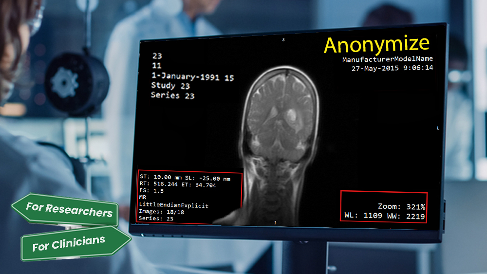
- Anonymized DICOM handling
- Role-based access controls
Trusted security — built for modern care.

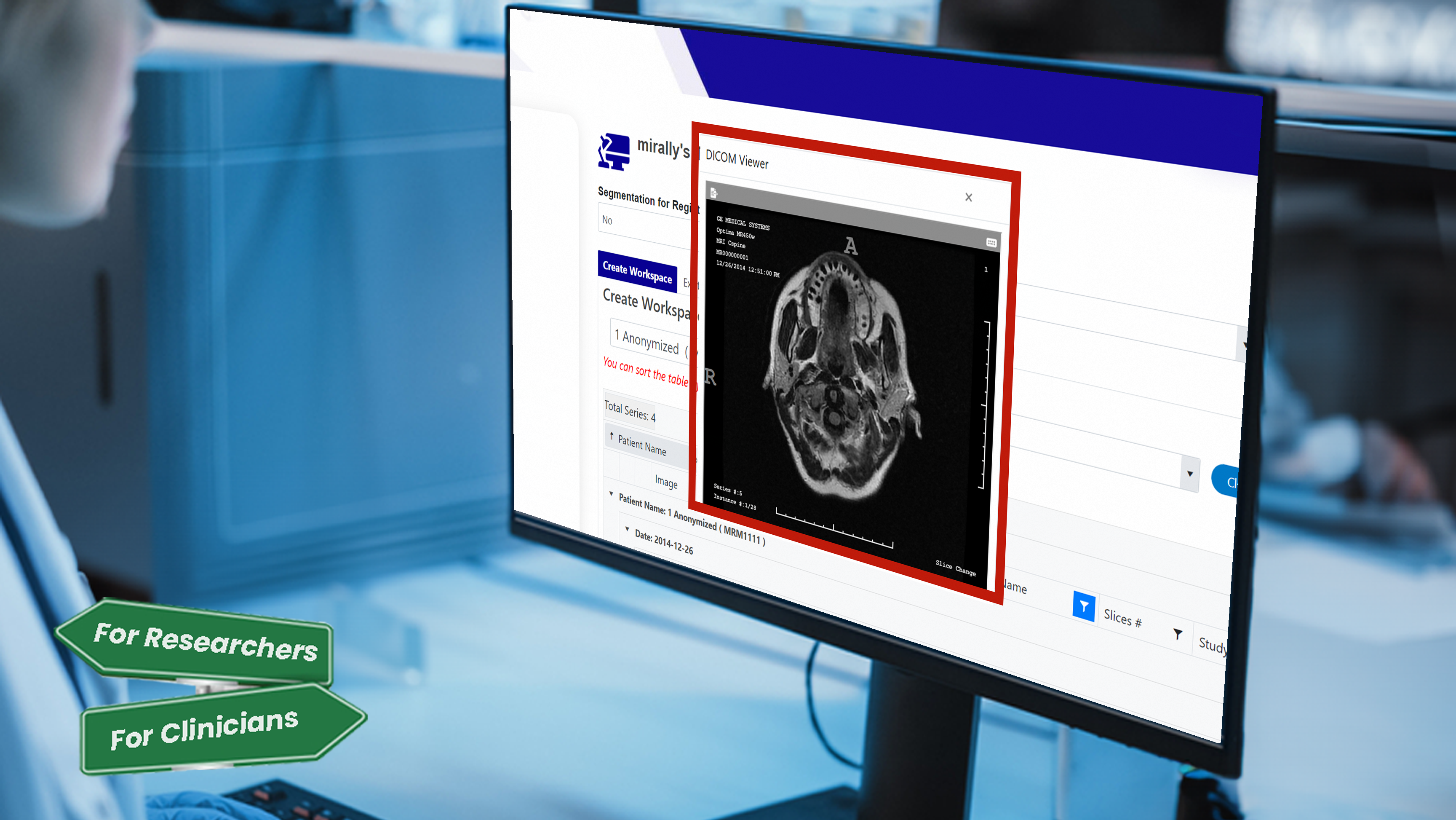
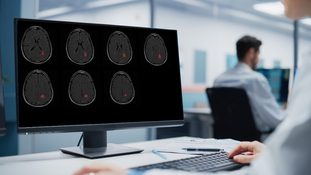
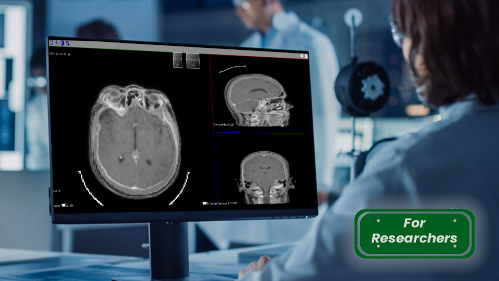
-GBM DICOM Viewer
High-resolution MRI viewing, purpose-built for glioblastoma.
Access and review magnetic resonance images (MRI) of glioblastoma patients directly in-browser using our FDA-cleared DICOM viewer. Designed for both clinical and research workflows, the tool enables fast navigation, accurate interpretation, and seamless integration with volumetric segmentation.
- Built-in GBM-focused overlays
- Slice-by-slice navigation
- Optimized for speed and clarity
-Download Segmentation
Flexible export options for clinical and research use.
Access and download AI-generated segmentations in the format that fits your workflow:
- RT-DICOM — optimized for radiotherapy planning (clinicians & researchers)
- Standard DICOM — for use across most clinical systems (clinicians & researchers)
- NIfTI — research-grade format for deep learning and analysis (researchers only)
FDA-cleared and compatible with most imaging pipelines.
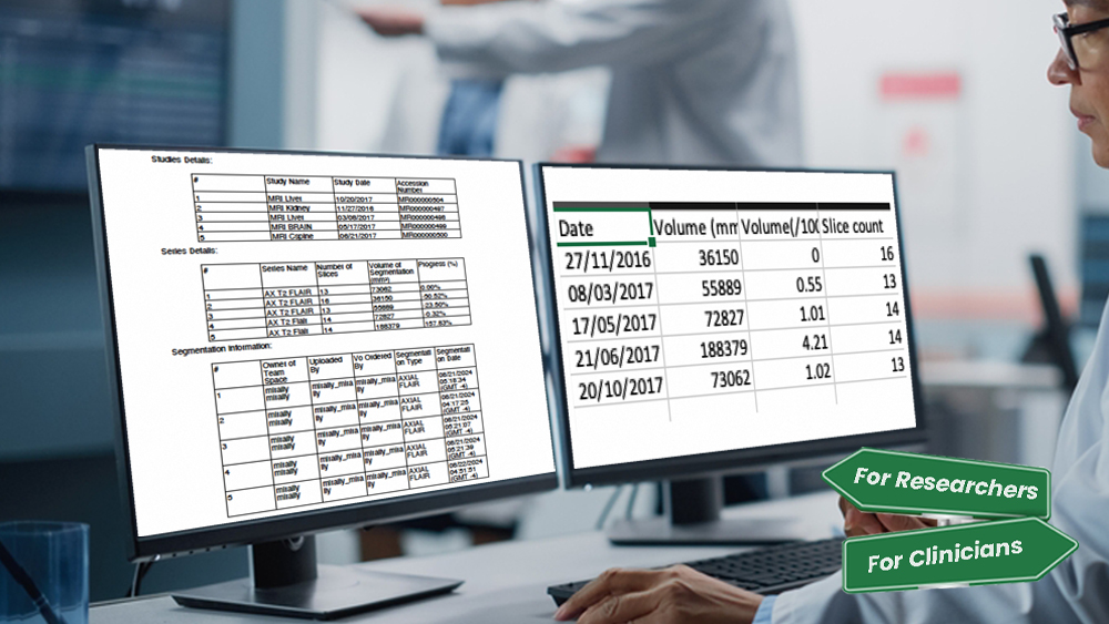
-Download Volumes
Quantitative outputs for clinical reports and research analysis.
Export tumor volume data in multiple formats to suit your workflow:
- Excel — for easy tracking, filtering, and cross-patient comparison
- PDF — clean, shareable reports for documentation and teams
- Plot visuals — fast insights into tumor progression over time
All outputs are traceable, standardized, and FDA-cleared.
-Registration
Seamless alignment across modalities, powered by AI.
MRIMath’s registration tools are built to streamline your workflow without compromising precision. Whether you’re working with MR, CT,
or multi-modal datasets, our platform enables fast and accurate image alignment tailored to clinical and research needs.
We support both:
- AI-powered rigid registration for the brain
- Software-assisted manual workflows for fine-tuned control
Designed for researchers. Trusted by clinicians.
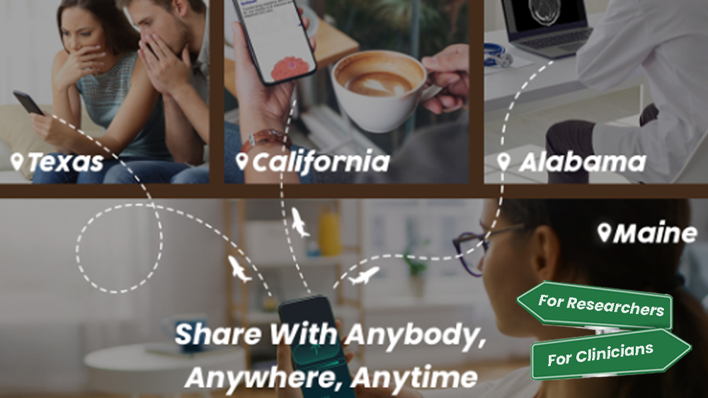
-Sharing & Exchange
Seamless access. No CDs. No delays.
Patients, physicians, and institutions can instantly share or access imaging studies from anywhere — without burning discs or relying on faxed forms. MRIMath’s exchange tools make data flow securely and efficiently, whether across states or across departments.- Share and receive DICOM files with one click
- Choose between anonymized or PHI-included transfers
- Set access permissions: temporary or permanent
Built for modern care. Designed to empower researchers and clinicians alike.
-Anonymization Service
Committed to Health. Committed to Privacy.
Secure. Fast. Fully compatible with clinical and research workflows.